

How Long Does It Take For The Liver To Regenerate
| Liver | |
|---|---|
 The human liver is located in the upper right abdomen | |
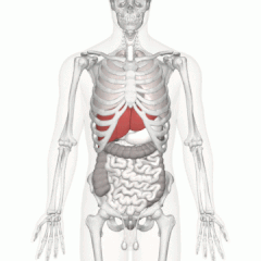 Location of homo liver (in reddish) shown on a male trunk | |
| Details | |
| Forerunner | Foregut |
| System | Digestive organisation |
| Avenue | Hepatic artery |
| Vein | Hepatic vein and hepatic portal vein |
| Nerve | Celiac ganglia and vagus nervus[1] |
| Identifiers | |
| Latin | Jecur, iecur |
| Greek | Hepar (ἧπαρ) root hepat- (ἡπατ-) |
| MeSH | D008099 |
| TA98 | A05.eight.01.001 |
| TA2 | 3023 |
| FMA | 7197 |
| Anatomical terminology [edit on Wikidata] | |
The liver is a major organ only found in vertebrates which performs many essential biological functions such as detoxification of the organism, and the synthesis of proteins and biochemicals necessary for digestion and growth.[two] [iii] [4] In humans, it is located in the right upper quadrant of the abdomen, below the diaphragm. Its other roles in metabolism include the regulation of glycogen storage, decomposition of red claret cells, and the product of hormones.[iv]
The liver is an accessory digestive organ that produces bile, an element of group i fluid containing cholesterol and bile acids, which helps the breakdown of fatty. The gallbladder, a small pouch that sits just under the liver, stores bile produced by the liver which is afterwards moved to the small intestine to consummate digestion.[v] The liver'south highly specialized tissue, consisting of generally hepatocytes, regulates a wide multifariousness of loftier-book biochemical reactions, including the synthesis and breakup of minor and complex molecules, many of which are necessary for normal vital functions.[six] Estimates regarding the organ'southward full number of functions vary, but textbooks mostly cite it being around 500.[7]
It is non known how to compensate for the absence of liver part in the long term, although liver dialysis techniques tin can be used in the short term. Bogus livers have not been adult to promote long-term replacement in the absenteeism of the liver. Every bit of 2018[update],[8] liver transplantation is the only option for complete liver failure.
Construction [edit]

The liver, viewed from in a higher place, showing the left and right lobes separated by the falciform ligament
The liver is a reddish-brown, wedge-shaped organ with two lobes of unequal size and shape. A human liver normally weighs approximately one.5 kg (three.iii lb)[ix] and has a width of about 15 cm (6 in).[x] There is considerable size variation between individuals, with the standard reference range for men being 970–1,860 g (2.14–4.10 lb)[xi] and for women 600–1,770 g (ane.32–3.90 lb).[12] It is both the heaviest internal organ and the largest gland in the human body. Located in the correct upper quadrant of the abdominal cavity, it rests just below the diaphragm, to the correct of the stomach and overlies the gallbladder.[5]
The liver is connected to two large claret vessels: the hepatic artery and the portal vein. The hepatic artery carries oxygen-rich blood from the aorta via the celiac trunk, whereas the portal vein carries blood rich in digested nutrients from the unabridged alimentary canal and also from the spleen and pancreas.[8] These blood vessels subdivide into minor capillaries known as liver sinusoids, which then lead to lobules.
Lobules are the functional units of the liver. Each lobule is fabricated upwards of millions of hepatic cells (hepatocytes), which are the basic metabolic cells. The lobules are held together past a fine, dense, irregular, fibroelastic connective tissue layer extending from the gristly capsule roofing the entire liver known equally Glisson'south capsule.[4] This extends into the structure of the liver past accompanying the blood vessels, ducts, and nerves at the hepatic hilum. The whole surface of the liver, except for the blank expanse, is covered in a serous coat derived from the peritoneum, and this firmly adheres to the inner Glisson'southward sheathing.
Gross beefcake [edit]
Terminology related to the liver frequently starts in hepat- from ἡπατο-, from the Greek word for liver.[13]
Lobes [edit]
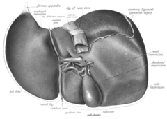
The liver, viewed from below, surface showing four lobes and the impressions
The liver is grossly divided into ii parts when viewed from above – a right and a left lobe - and four parts when viewed from below (left, right, caudate, and quadrate lobes).[xiv]
The falciform ligament makes a superficial division of the liver into a left and right lobe. From below, the two additional lobes are located between the right and left lobes, one in front of the other. A line tin can be imagined running from the left of the vena cava and all the way forward to dissever the liver and gallbladder into two halves.[15] This line is called Cantlie'southward line.[16]
Other anatomical landmarks include the ligamentum venosum and the round ligament of the liver, which further separate the left side of the liver in 2 sections. An important anatomical landmark, the porta hepatis, divides this left portion into four segments, which can be numbered starting at the caudate lobe as I in an anticlockwise manner. From this parietal view, seven segments can be seen, because the eighth segment is simply visible in the visceral view.[17]
Surfaces [edit]
On the diaphragmatic surface, autonomously from a triangular blank area where it connects to the diaphragm, the liver is covered by a sparse, double-layered membrane, the peritoneum, that helps to reduce friction against other organs.[18] This surface covers the convex shape of the two lobes where it accommodates the shape of the diaphragm. The peritoneum folds back on itself to form the falciform ligament and the right and left triangular ligaments.[19]
These peritoneal ligaments are not related to the anatomic ligaments in joints, and the correct and left triangular ligaments have no known functional importance, though they serve as surface landmarks.[19] The falciform ligament functions to attach the liver to the posterior portion of the anterior trunk wall.
The visceral surface or inferior surface is uneven and concave. It is covered in peritoneum apart from where it attaches the gallbladder and the porta hepatis.[xviii] The fossa of gall float lies to the right of the quadrate lobe, occupied by the gallbladder with its cystic duct close to the correct end of porta hepatis.
Impressions [edit]

Several impressions on the surface of the liver adapt the various side by side structures and organs. Underneath the correct lobe and to the correct of the gallbladder fossa are two impressions, one backside the other and separated past a ridge. The ane in front end is a shallow colic impression, formed by the hepatic flexure and the ane behind is a deeper renal impression accommodating part of the right kidney and part of the suprarenal gland.[20]
The suprarenal impression is a small, triangular, depressed area on the liver. It is located close to the correct of the fossa, between the bare expanse and the caudate lobe, and immediately higher up the renal impression. The greater role of the suprarenal impression is devoid of peritoneum and it lodges the right suprarenal gland.[21]
Medial to the renal impression is a tertiary and slightly marked impression, lying between it and the neck of the gall float. This is caused by the descending portion of the duodenum, and is known as the duodenal impression.[21]
The inferior surface of the left lobe of the liver presents behind and to the left of the gastric impression.[21] This is moulded over the upper front end surface of the tummy, and to the right of this is a rounded eminence, the tuber omentale, which fits into the concavity of the lesser curvature of the stomach and lies in forepart of the anterior layer of the lesser omentum.
Microscopic beefcake [edit]

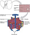
Cells, ducts, and blood vessels
Microscopically, each liver lobe is seen to be made up of hepatic lobules. The lobules are roughly hexagonal, and consist of plates of hepatocytes, and sinusoids radiating from a central vein towards an imaginary perimeter of interlobular portal triads.[22] The central vein joins to the hepatic vein to carry blood out from the liver. A distinctive component of a lobule is the portal triad, which can be found running along each of the lobule's corners. The portal triad, consists of the hepatic artery, the portal vein, and the common bile duct.[23] The triad may be seen on a liver ultrasound, as a Mickey Mouse sign with the portal vein as the head, and the hepatic artery, and the common bile duct equally the ears.[24]
Histology, the study of microscopic anatomy, shows two major types of liver cell: parenchymal cells and nonparenchymal cells. Almost 70–85% of the liver volume is occupied past parenchymal hepatocytes. Nonparenchymal cells plant 40% of the total number of liver cells just but six.5% of its volume.[25] The liver sinusoids are lined with two types of cell, sinusoidal endothelial cells, and phagocytic Kupffer cells.[26] Hepatic stellate cells are nonparenchymal cells establish in the perisinusoidal space, between a sinusoid and a hepatocyte.[25] Additionally, intrahepatic lymphocytes are often present in the sinusoidal lumen.[25]
-
Microscopic anatomy of the liver
-

Types of capillaries–sinusoid on right
-

3D Medical Animation Still Shot Depicting parts of liver
Functional anatomy [edit]
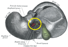
Hilum of the liver, circled in yellow
The central area or hepatic hilum, includes the opening known as the porta hepatis which carries the common bile duct and common hepatic avenue, and the opening for the portal vein. The duct, vein, and artery dissever into left and right branches, and the areas of the liver supplied by these branches constitute the functional left and correct lobes. The functional lobes are separated by the imaginary plane, Cantlie's line, joining the gallbladder fossa to the inferior vena cava. The airplane separates the liver into the truthful right and left lobes. The middle hepatic vein also demarcates the true right and left lobes. The right lobe is further divided into an anterior and posterior segment by the right hepatic vein. The left lobe is divided into the medial and lateral segments by the left hepatic vein.
The hilum of the liver is described in terms of three plates that contain the bile ducts and blood vessels. The contents of the whole plate arrangement are surrounded by a sheath.[27] The three plates are the hilar plate, the cystic plate and the umbilical plate and the plate organization is the site of the many anatomical variations to be constitute in the liver.[27]
Couinaud classification organization [edit]

Shape of homo liver in animation, with eight Couinaud segments labelled
In the widely used Couinaud organization, the functional lobes are farther divided into a full of eight subsegments based on a transverse airplane through the bifurcation of the master portal vein.[28] The caudate lobe is a dissever structure that receives claret flow from both the right- and left-sided vascular branches.[29] [30] The Couinaud classification divides the liver into eight functionally contained liver segments. Each segment has its own vascular inflow, outflow and biliary drainage. In the centre of each segment are branches of the portal vein, hepatic avenue, and bile duct. In the periphery of each segment is vascular outflow through the hepatic veins.[31] The nomenclature system uses the vascular supply in the liver to separate the functional units (numbered I to Eight) with unit of measurement 1, the caudate lobe, receiving its supply from both the right and the left branches of the portal vein. It contains one or more hepatic veins which drain straight into the inferior vena cava.[28] The remainder of the units (2 to Viii) are numbered in a clockwise manner:[31]
Cistron and poly peptide expression [edit]
About 20,000 poly peptide coding genes are expressed in human cells and lx% of these genes are expressed in a normal, adult liver.[32] [33] Over 400 genes are more than specifically expressed in the liver, with some 150 genes highly specific for liver tissue. A large fraction of the respective liver specific proteins are mainly expressed in hepatocytes and secreted into the blood and found plasma proteins. Other liver specific proteins are sure liver enzymes such as HAO1 and RDH16, proteins involved in bile synthesis such every bit BAAT and SLC27A5, and transporter proteins involved in the metabolism of drugs, such as ABCB11 and SLC2A2. Examples of highly liver-specific proteins include apolipoprotein A 2, coagulation factors F2 and F9, complement factor related proteins, and the fibrinogen beta chain poly peptide.[34]
Development [edit]

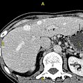
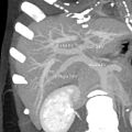
CT scan showing an adult liver in the axial plane.
Organogenesis, the development of the organs, takes place from the third to the eighth calendar week during embryogenesis. The origins of the liver prevarication in both the ventral portion of the foregut endoderm (endoderm being one of the iii embryonic germ layers) and the constituents of the adjacent septum transversum mesenchyme. In the homo embryo, the hepatic diverticulum is the tube of endoderm that extends out from the foregut into the surrounding mesenchyme. The mesenchyme of septum transversum induces this endoderm to proliferate, to branch, and to form the glandular epithelium of the liver. A portion of the hepatic diverticulum (that region closest to the digestive tube) continues to function as the drainage duct of the liver, and a branch from this duct produces the gallbladder.[35] Also signals from the septum transversum mesenchyme, fibroblast growth factor from the developing heart too contributes to hepatic competence, along with retinoic acrid emanating from the lateral plate mesoderm. The hepatic endodermal cells undergo a morphological transition from columnar to pseudostratified resulting in thickening into the early on liver bud. Their expansion forms a population of the bipotential hepatoblasts.[36] Hepatic stellate cells are derived from mesenchyme.[37]
After migration of hepatoblasts into the septum transversum mesenchyme, the hepatic architecture begins to be established, with liver sinusoids and bile canaliculi appearing. The liver bud separates into the lobes. The left umbilical vein becomes the ductus venosus and the correct vitelline vein becomes the portal vein. The expanding liver bud is colonized by hematopoietic cells. The bipotential hepatoblasts begin differentiating into biliary epithelial cells and hepatocytes. The biliary epithelial cells differentiate from hepatoblasts around portal veins, first producing a monolayer, and so a bilayer of cuboidal cells. In ductal plate, focal dilations emerge at points in the bilayer, get surrounded by portal mesenchyme, and undergo tubulogenesis into intrahepatic bile ducts. Hepatoblasts not adjacent to portal veins instead differentiate into hepatocytes and arrange into cords lined by sinusoidal epithelial cells and bile canaliculi. In one case hepatoblasts are specified into hepatocytes and undergo farther expansion, they begin acquiring the functions of a mature hepatocyte, and eventually mature hepatocytes appear every bit highly polarized epithelial cells with abundant glycogen accumulation. In the adult liver, hepatocytes are not equivalent, with position forth the portocentrovenular axis within a liver lobule dictating expression of metabolic genes involved in drug metabolism, carbohydrate metabolism, ammonia detoxification, and bile product and secretion. WNT/β-catenin has at present been identified to exist playing a key role in this miracle.[36]
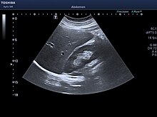
Adult ultrasound showing the correct lobe of the liver and right kidney.
At birth, the liver comprises roughly 4% of torso weight and weighs on average about 120 g (4 oz). Over the grade of further development, it volition increase to 1.4–1.half-dozen kg (3.one–3.5 lb) just will only take upward 2.five–3.5% of torso weight.[38]
Fetal claret supply [edit]
In the growing fetus, a major source of blood to the liver is the umbilical vein, which supplies nutrients to the growing fetus. The umbilical vein enters the abdomen at the navel and passes upward forth the complimentary margin of the falciform ligament of the liver to the junior surface of the liver. There, information technology joins with the left branch of the portal vein. The ductus venosus carries blood from the left portal vein to the left hepatic vein and then to the inferior vena cava, allowing placental blood to bypass the liver. In the fetus, the liver does not perform the normal digestive processes and filtration of the infant liver considering nutrients are received directly from the female parent via the placenta. The fetal liver releases some blood stem cells that migrate to the fetal thymus, creating the T-cells or T-lymphocytes. Afterwards birth, the formation of blood stalk cells shifts to the reddish bone marrow. After ii–5 days, the umbilical vein and ductus venosus are obliterated; the former becomes the circular ligament of liver and the latter becomes the ligamentum venosum. In the disorders of cirrhosis and portal hypertension, the umbilical vein can open up again.
Functions [edit]
The various functions of the liver are carried out past the liver cells or hepatocytes. The liver is thought to be responsible for upwardly to 500 separate functions, ordinarily in combination with other systems and organs. Currently, no artificial organ or device is capable of reproducing all the functions of the liver. Some functions can exist carried out by liver dialysis, an experimental treatment for liver failure. The liver too accounts for about xx% of resting full body oxygen consumption.
Claret supply [edit]
The liver receives a dual blood supply from the hepatic portal vein and hepatic arteries. The hepatic portal vein delivers around 75% of the liver's blood supply and carries venous blood drained from the spleen, gastrointestinal tract, and its associated organs. The hepatic arteries supply arterial blood to the liver, accounting for the remaining quarter of its blood flow. Oxygen is provided from both sources; about half of the liver's oxygen demand is met past the hepatic portal vein, and half is met by the hepatic arteries.[39] The hepatic artery also has both alpha- and beta-adrenergic receptors; therefore, flow through the artery is controlled, in office, by the splanchnic nerves of the autonomic nervous arrangement.
Claret flows through the liver sinusoids and empties into the central vein of each lobule. The fundamental veins coalesce into hepatic veins, which leave the liver and drain into the inferior vena cava.[40]
-

Liver veins
-

Diagram of liver, lobule, and portal tract and their inter-relations
Biliary menstruation [edit]

The biliary tract is derived from the branches of the bile ducts. The biliary tract, likewise known as the biliary tree, is the path by which bile is secreted by the liver then transported to the first part of the small intestine, the duodenum. The bile produced in the liver is collected in bile canaliculi, minor grooves between the faces of next hepatocytes. The canaliculi radiate to the edge of the liver lobule, where they merge to form bile ducts. Within the liver, these ducts are termed intrahepatic bile ducts, and once they exit the liver, they are considered extrahepatic. The intrahepatic ducts eventually drain into the right and left hepatic ducts, which exit the liver at the transverse fissure, and merge to grade the common hepatic duct. The cystic duct from the gallbladder joins with the common hepatic duct to course the mutual bile duct.[xl] The biliary system and connective tissue is supplied by the hepatic avenue alone.
Bile either drains directly into the duodenum via the common bile duct, or is temporarily stored in the gallbladder via the cystic duct. The mutual bile duct and the pancreatic duct enter the second function of the duodenum together at the hepatopancreatic ampulla, also known as the ampulla of Vater.
Metabolism [edit]
The liver plays a major role in carbohydrate, poly peptide, amino acrid, and lipid metabolism.
Carbohydrate metabolism [edit]
The liver performs several roles in saccharide metabolism:
- The liver synthesizes and stores around 100g of glycogen via glycogenesis, the germination of glycogen from glucose.
- When needed, the liver releases glucose into the blood past performing glycogenolysis, the breakdown of glycogen into glucose.[41]
- The liver is also responsible for gluconeogenesis, which is the synthesis of glucose from certain amino acids, lactate, or glycerol. Adipose and liver cells produce glycerol past breakdown of fat, which the liver uses for gluconeogenesis.[41]
- Liver also does glyconeogenesis which is synthesis of glycogen from lactic acid.[42]
Protein metabolism [edit]
The liver is responsible for the mainstay of protein metabolism, synthesis equally well as degradation. All plasma proteins except Gamma-globulins are synthesised in the liver.[43] Information technology is also responsible for a large part of amino acid synthesis. The liver plays a role in the production of clotting factors, every bit well equally red blood cell product. Some of the proteins synthesized by the liver include coagulation factors I (fibrinogen), Two (prothrombin), V, VII, VIII, Nine, X, XI, XII, Thirteen, as well as protein C, protein S and antithrombin. The liver is a major site of production for thrombopoietin, a glycoprotein hormone that regulates the production of platelets by the bone marrow.[44]
Lipid metabolism [edit]
The liver plays several roles in lipid metabolism: information technology performs cholesterol synthesis, lipogenesis, and the production of triglycerides, and a majority of the body'southward lipoproteins are synthesized in the liver. The liver plays a key role in digestion, as it produces and excretes bile (a yellowish liquid) required for emulsifying fats and help the absorption of vitamin K from the diet. Some of the bile drains straight into the duodenum, and some is stored in the gallbladder. The liver produces insulin-like growth cistron 1, a polypeptide poly peptide hormone that plays an important function in childhood growth and continues to have anabolic effects in adults.
Breakup [edit]
The liver is responsible for the breakdown of insulin and other hormones. The liver breaks downwardly bilirubin via glucuronidation, facilitating its excretion into bile. The liver is responsible for the breakdown and excretion of many waste product products. Information technology plays a cardinal office in breaking down or modifying toxic substances (e.g., methylation) and most medicinal products in a procedure called drug metabolism. This sometimes results in toxication, when the metabolite is more toxic than its precursor. Preferably, the toxins are conjugated to avail excretion in bile or urine. The liver converts ammonia into urea as part of the ornithine cycle or the urea cycle, and the urea is excreted in the urine.[45]
Blood reservoir [edit]
Because the liver is an expandable organ, large quantities of blood can exist stored in its blood vessels. Its normal claret volume, including both that in the hepatic veins and that in the hepatic sinuses, is almost 450 milliliters, or virtually 10 percent of the body's full blood volume. When high pressure in the right atrium causes backpressure in the liver, the liver expands, and 0.5 to 1 liter of extra blood is occasionally stored in the hepatic veins and sinuses. This occurs especially in cardiac failure with peripheral congestion. Thus, in effect, the liver is a large, expandable, venous organ capable of acting as a valuable blood reservoir in times of excess claret volume and capable of supplying extra blood in times of macerated blood volume.[46]
Lymph production [edit]
Because the pores in the hepatic sinusoids are very permeable and allow ready passage of both fluid and proteins into the perisinusoidal space, the lymph draining from the liver usually has a protein concentration of well-nigh 6 g/dl, which is only slightly less than the protein concentration of plasma. Also, the high permeability of the liver sinusoid epithelium allows big quantities of lymph to form. Therefore, almost half of all the lymph formed in the body under resting conditions arises in the liver.
Other [edit]
- The liver stores a multitude of substances, including vitamin A (1–2 years' supply), vitamin D (ane–4 months' supply),[47] vitamin B12 (iii–5 years' supply),[48] vitamin K, vitamin East, iron, copper, zinc, cobalt, molybdenum, etc.
- Haemopoiesis - The formation of blood cells is called haemopoiesis. In embryonic stage RBC and WBC are formed past liver. In the first trimester fetus, the liver is the main site of red blood cell production. Past the 32nd week of gestation, the bone marrow has almost completely taken over that task.[49]
- Liver helps in purification of blood. The Kupffer cells of liver are phagocytic cells, helps in phagocytosis of dead blood cells and bacteria from the blood.[l]
- The liver is responsible for immunological effects – the mononuclear phagocyte arrangement of the liver contains many immunologically active cells, acting as a 'sieve' for antigens carried to it via the portal system.
- The liver produces albumin, the most arable protein in claret serum. It is essential in the maintenance of oncotic pressure, and acts as a ship for fat acids and steroid hormones.
- The liver synthesizes angiotensinogen, a hormone that is responsible for raising the blood pressure level when activated by renin, an enzyme that is released when the kidney senses depression claret pressure.
- The liver produces the enzyme catalase to intermission down hydrogen peroxide, a toxic oxidising agent, into water and oxygen.
With crumbling [edit]
The oxidative capacity of the liver decreases with aging, and therefore any medications that crave oxidation (for instance, benzodiazepines) are more likely to accumulate to toxic levels. Even so, medications with shorter half-lives, such as lorazepam and oxazepam, are preferred in most cases when benzodiazepines are required in regard to geriatric medicine.
Clinical significance [edit]
Disease [edit]
The liver is a vital organ and supports nearly every other organ in the body. Considering of its strategic location and multidimensional functions, the liver is also prone to many diseases.[51] The bare area of the liver is a site that is vulnerable to the passing of infection from the abdominal cavity to the thoracic crenel. Liver diseases may be diagnosed by liver function tests–blood tests that tin identify various markers. For case, acute-stage reactants are produced by the liver in response to injury or inflammation.
Hepatitis is a common condition of inflammation of the liver. The most usual crusade of this is viral, and the most common of these infections are hepatitis A, B, C, D, and E. Some of these infections are sexually transmitted. Inflammation can also be caused by other viruses in the family Herpesviridae such as the herpes simplex virus. Chronic (rather than acute) infection with hepatitis B virus or hepatitis C virus is the principal crusade of liver cancer.[52] Globally, about 248 million individuals are chronically infected with hepatitis B (with 843,724 in the U.Southward.),[53] and 142 million are chronically infected with hepatitis C[54] (with 2.7 million in the U.Southward.[55]). Globally at that place are about 114 million and twenty meg cases of hepatitis A[54] and hepatitis E[56] respectively, but these generally resolve and do not go chronic. Hepatitis D virus is a "satellite" of hepatitis B virus (tin only infect in the presence of hepatitis B), and co-infects nearly 20 1000000 people with hepatitis B, globally.[57]
Hepatic encephalopathy is caused by an aggregating of toxins in the bloodstream that are normally removed by the liver. This condition can result in coma and tin prove fatal. Budd–Chiari syndrome is a condition caused past blockage of the hepatic veins (including thrombosis) that drain the liver. Information technology presents with the classical triad of abdominal pain, ascites and liver enlargement.[58] Many diseases of the liver are accompanied by jaundice caused past increased levels of bilirubin in the system. The bilirubin results from the breakdown of the hemoglobin of dead red blood cells; normally, the liver removes bilirubin from the blood and excretes it through bile.
Other disorders caused past excessive alcohol consumption are grouped under alcoholic liver diseases and these include alcoholic hepatitis, fatty liver, and cirrhosis. Factors contributing to the evolution of alcoholic liver diseases are not only the quantity and frequency of alcohol consumption, only can also include gender, genetics, and liver insult. Liver damage can likewise be acquired by drugs, particularly paracetamol and drugs used to treat cancer. A rupture of the liver can be caused past a liver shot used in gainsay sports.
Chief biliary cholangitis is an autoimmune affliction of the liver.[59] [60] Information technology is marked by irksome progressive destruction of the small bile ducts of the liver, with the intralobular ducts (Canals of Hering) affected early on in the disease.[61] When these ducts are damaged, bile and other toxins build up in the liver (cholestasis) and over time damages the liver tissue in combination with ongoing allowed related impairment. This can lead to scarring (fibrosis) and cirrhosis. Cirrhosis increases the resistance to blood menstruum in the liver, and can effect in portal hypertension. Congested anastomoses betwixt the portal venous system and the systemic apportionment, tin can exist a subsequent condition.
In that location are likewise many pediatric liver diseases, including biliary atresia, alpha-ane antitrypsin deficiency, alagille syndrome, progressive familial intrahepatic cholestasis, Langerhans cell histiocytosis and hepatic hemangioma a benign tumour the virtually common blazon of liver tumour, idea to exist congenital. A genetic disorder causing multiple cysts to form in the liver tissue, usually in afterward life, and ordinarily asymptomatic, is polycystic liver disease. Diseases that interfere with liver function will atomic number 82 to derangement of these processes. However, the liver has a great capacity to regenerate and has a big reserve chapters. In nigh cases, the liver only produces symptoms after extensive harm.
Hepatomegaly refers to an enlarged liver and tin can be due to many causes. Information technology tin be palpated in a liver span measurement.
Symptoms [edit]
The classic symptoms of liver damage include the following:
- Pale stools occur when stercobilin, a brown pigment, is absent from the stool. Stercobilin is derived from bilirubin metabolites produced in the liver.
- Dark urine occurs when bilirubin mixes with urine
- Jaundice (yellow skin and/or whites of the eyes) This is where bilirubin deposits in skin, causing an intense itch. Itching is the near common complaint by people who have liver failure. Ofttimes this itch cannot exist relieved by drugs.
- Swelling of the belly, and swelling of the ankles and feet occurs because the liver fails to make albumin.
- Excessive fatigue occurs from a generalized loss of nutrients, minerals and vitamins.
- Bruising and easy bleeding are other features of liver disease. The liver makes clotting factors, substances which aid prevent bleeding. When liver damage occurs, these factors are no longer present and severe bleeding can occur.[62]
- Pain in the upper right quadrant tin consequence from the stretching of Glisson'southward capsule in weather of hepatitis and pre-eclampsia.
Diagnosis [edit]
The diagnosis of liver disease is made by liver function tests, groups of claret tests, that can readily evidence the extent of liver harm. If infection is suspected, then other serological tests will be carried out. A physical test of the liver can simply reveal its size and any tenderness, and some form of imaging such as an ultrasound or CT scan may besides be needed.[63] Sometimes a liver biopsy will exist necessary, and a tissue sample is taken through a needle inserted into the skin simply beneath the rib muzzle. This process may be helped by a sonographer providing ultrasound guidance to an interventional radiologist.[64]
-
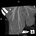
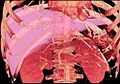
Axial CT prototype showing anomalous hepatic veins coursing on the subcapsular anterior surface of the liver.[65]
-
Maximum intensity projection (MIP) CT image as viewed anteriorly showing the dissonant hepatic veins coursing on the inductive surface of the liver
-
Lateral MIP view in the same patient
-

A CT scan in which the liver and portal vein are shown.
Liver regeneration [edit]
The liver is the simply man internal organ capable of natural regeneration of lost tissue; as little as 25% of a liver tin regenerate into a whole liver.[66] This is, however, not true regeneration simply rather compensatory growth in mammals.[67] The lobes that are removed practice not regrow and the growth of the liver is a restoration of part, not original form. This contrasts with true regeneration where both original function and class are restored. In some other species, such every bit zebrafish, the liver undergoes truthful regeneration by restoring both shape and size of the organ.[68] In the liver, big areas of the tissues are formed but for the formation of new cells there must be sufficient corporeality of material then the apportionment of the claret becomes more active.[69]
This is predominantly due to the hepatocytes re-entering the cell wheel. That is, the hepatocytes go from the quiescent G0 phase to the G1 stage and undergo mitosis. This procedure is activated past the p75 receptors.[lxx] There is also some show of bipotential stem cells, called hepatic oval cells or ovalocytes (not to be confused with oval reddish blood cells of ovalocytosis), which are thought to reside in the canals of Hering. These cells can differentiate into either hepatocytes or cholangiocytes. Cholangiocytes are the epithelial lining cells of the bile ducts.[71] They are cuboidal epithelium in the minor interlobular bile ducts, but become columnar and mucus secreting in larger bile ducts approaching the porta hepatis and the extrahepatic ducts. Research is being carried out on the employ of stem cells for the generation of an bogus liver.
Scientific and medical works about liver regeneration often refer to the Greek Titan Prometheus who was chained to a stone in the Caucasus where, each day, his liver was devoured by an hawkeye, simply to grow back each night. The myth suggests the ancient Greeks may have known most the liver'southward remarkable chapters for self-repair.[72]
Liver transplantation [edit]
Human liver transplants were starting time performed by Thomas Starzl in the United States and Roy Calne in Cambridge, England in 1963 and 1967, respectively.
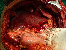
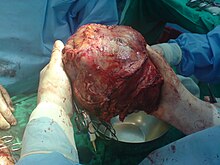
After resection of left lobe liver tumor
Liver transplantation is the but option for those with irreversible liver failure. Most transplants are washed for chronic liver diseases leading to cirrhosis, such as chronic hepatitis C, alcoholism, and autoimmune hepatitis. Less commonly, liver transplantation is washed for fulminant hepatic failure, in which liver failure occurs over days to weeks.
Liver allografts for transplant usually come up from donors who take died from fatal brain injury. Living donor liver transplantation is a technique in which a portion of a living person'south liver is removed (hepatectomy) and used to replace the unabridged liver of the recipient. This was first performed in 1989 for pediatric liver transplantation. Only 20 percent of an adult's liver (Couinaud segments ii and 3) is needed to serve as a liver allograft for an infant or small kid.
More recently,[ when? ] adult-to-adult liver transplantation has been washed using the donor'south right hepatic lobe, which amounts to sixty percent of the liver. Due to the ability of the liver to regenerate, both the donor and recipient stop upwards with normal liver function if all goes well. This procedure is more controversial, equally it entails performing a much larger operation on the donor, and indeed there were at least ii donor deaths out of the outset several hundred cases. A 2006 publication addressed the problem of donor mortality and found at to the lowest degree fourteen cases.[73] The run a risk of postoperative complications (and expiry) is far greater in correct-sided operations than that in left-sided operations.
With the recent advances of noninvasive imaging, living liver donors usually accept to undergo imaging examinations for liver beefcake to decide if the anatomy is feasible for donation. The evaluation is normally performed past multidetector row computed tomography (MDCT) and magnetic resonance imaging (MRI). MDCT is expert in vascular anatomy and volumetry. MRI is used for biliary tree anatomy. Donors with very unusual vascular anatomy, which makes them unsuitable for donation, could be screened out to avert unnecessary operations.
-

MDCT image. Arterial anatomy contraindicated for liver donation
-
MDCT paradigm. Portal venous beefcake contraindicated for liver donation
-
MDCT image. 3D image created by MDCT can clearly visualize the liver, mensurate the liver volume, and plan the dissection aeroplane to facilitate the liver transplantation procedure.
-

Phase contrast CT image. Contrast is perfusing the right liver only non the left due to a left portal vein thrombus.
Gild and culture [edit]
Some cultures regard the liver as the seat of the soul.[74] In Greek mythology, the gods punished Prometheus for revealing fire to humans past chaining him to a rock where a vulture (or an eagle) would peck out his liver, which would regenerate overnight. (The liver is the only human internal organ that actually can regenerate itself to a meaning extent.) Many ancient peoples of the Nearly Eastward and Mediterranean areas practiced a type of divination called haruspicy or hepatomancy, where they tried to obtain information by examining the livers of sheep and other animals.
In Plato, and in later physiology, the liver was thought to be the seat of the darkest emotions (specifically wrath, jealousy and greed) which bulldoze men to action.[75] The Talmud (tractate Berakhot 61b) refers to the liver equally the seat of anger, with the gallbladder counteracting this. The Farsi, Urdu, and Hindi languages ( جگر or जिगर or jigar ) refer to the liver in figurative speech to indicate courage and stiff feelings, or "their best"; eastward.m., "This Mecca has thrown to yous the pieces of its liver!".[76] The term jan e jigar , literally "the strength (power) of my liver", is a term of endearment in Urdu. In Persian slang, jigar is used as an adjective for any object which is desirable, especially women. In the Zulu language, the word for liver ( isibindi ) is the same as the word for courage. In English the term 'lily-livered' is used to point cowardice from the medieval belief that the liver was the seat of courage. Castilian hígados also means "courage".[77] Nonetheless the secondary significant of Basque gibel is "indolence".[78]
In biblical Hebrew, the word for liver, כבד ( Kauved , stemmed KBD or KVD , similar to Arabic الكبد ), also means heavy and is used to describe the rich ("heavy" with possessions) and award (presumably for the aforementioned reason). In the Book of Lamentations (2:eleven) it is used to describe the physiological responses to sadness past "my liver spilled to globe" along with the flow of tears and the overturning in bitterness of the intestines.[79] On several occasions in the book of Psalms (most notably sixteen:9), the word is used to describe happiness in the liver, forth with the heart (which beats chop-chop) and the flesh (which appears cherry nether the skin). Farther usage as the self (like to "your honour") is widely available throughout the former testament, sometimes compared to the breathing soul (Genesis 49:6, Psalms vii:6, etc.). An honorable hat was besides referred to with this word (Task 19:ix, etc.) and under that definition appears many times along with פאר Pe'er - grandeur.[80]
These four meanings were used in preceding ancient Afro-Asiatic languages such equally Akkadian and Ancient Egyptian preserved in classical Ethiopic Ge'ez linguistic communication.[81]
Nutrient [edit]

Humans commonly eat the livers of mammals, fowl, and fish as food. Domestic pig, ox, lamb, dogie, chicken, and goose livers are widely available from butchers and supermarkets. In the Romance languages, the anatomical word for "liver" (French foie, Spanish hígado, etc.) derives non from the Latin anatomical term, jecur, only from the culinary term ficatum, literally "blimp with figs," referring to the livers of geese that had been fattened on figs.[82] Animal livers are rich in iron, vitamin A and vitamin B12; and cod liver oil is commonly used every bit a dietary supplement.
Liver can be baked, boiled, broiled, fried, stir-fried, or eaten raw (asbeh nayeh or sawda naye in Lebanese cuisine, or liver sashimi in Japanese cuisine). In many preparations, pieces of liver are combined with pieces of meat or kidneys, as in the various forms of Center Eastern mixed grill (e.yard. meurav Yerushalmi). Well-known examples include liver pâté, foie gras, chopped liver, and leverpastej. Liver sausages, such as Braunschweiger and liverwurst, are as well a valued meal. Liver sausages may likewise be used as spreads. A traditional South African delicacy, skilpadjies, is made of minced lamb's liver wrapped in netvet (caul fat), and grilled over an open fire. Traditionally, some fish livers were valued every bit nutrient, specially the stingray liver. It was used to set up delicacies, such as poached skate liver on toast in England, as well as the beignets de foie de raie and foie de raie en croute in French cuisine.[83]
Giraffe liver [edit]

The Humr, one of the tribes in the Baggara ethnic group, native to southwestern Kordofan in Sudan and speakers of Shuwa or Chadian Standard arabic, set up a non-alcoholic drink from the liver and bone marrow of the giraffe which they call umm nyolokh, and which they claim is intoxicating ( Standard arabic سكران sakran ), causing dreams and even waking hallucinations.[84] Anthropologist Ian Cunnison, who accompanied the Humr on ane of their giraffe-hunting expeditions in the late 1950s, notes that:
Information technology is said that a person, once he has drunk umm nyolokh, volition return to giraffe again and once again. Humr, being Mahdists, are strict abstainers [ from alcohol ] and a Humrawi is never drunkard ( sakran ) on liquor or beer. But he uses this discussion to describe the effects which umm nyolokh has upon him.[85]
Cunnison's remarkable account of an obviously psychoactive mammal found its manner from a somewhat obscure scientific newspaper into more than mainstream literature through a conversation between Dr. Wendy James of the Found of Social and Cultural Anthropology at the University of Oxford and specialist on the apply of hallucinogens and intoxicants in society Richard Rudgley, who considered its implications in his popular work The Encyclopedia of Psychoactive Substances. Rudgley hypothesises that the presence of the hallucinogenic chemical compound DMT might account for the putative intoxicating properties of umm nyolokh.[84]
Cunnison himself, on the other hand, had found it difficult fully to believe in the literal truth of the Humr's assertion that their drink was intoxicating:
I tin only assume that there is no exhilarant substance in the drinkable and that the effect it produces is but a thing of convention, although information technology may be brought almost subconsciously.[85]
The study of entheogens in general - including entheogens of animal origin ( e.one thousand. hallucinogenic fish and toad venom ) - has, nonetheless, made considerable progress in the lx-odd years since Cunnison'south report and the idea that some exhilarant principle might reside in giraffe liver no longer seems as far-fetched as it was in Cunnison's day, although conclusive proof ( or disproof ) will have to expect detailed analyses of the animal organ in question and the drink prepared therefrom.[84]
Arrow/bullet toxicant [edit]
Certain Tungusic peoples formerly prepared a blazon of pointer poison from rotting animal livers, which was, in later times, too applied to bullets. Russian anthropologist Sergei Mikhailovich Shirokogorov notes that:
Formerly the using of poisoned arrows was mutual. For example, amongst the Kumarčen, [ a subgroup of the Oroqen ] even in recent times a toxicant was used which was prepared from decomposable liver. * ( Note ) This has been confirmed by the Kumarčen. I am non competent to judge as to the chemical conditions of production of poison which is not destroyed by the heat of explosion. However, the Tungus themselves compare this method [ of poisoning ammunition ] with the poisoning of arrows.[86]
Other animals [edit]

The liver is found in all vertebrates and is typically the largest internal organ. Its form varies considerably in different species, and is largely determined by the shape and arrangement of the surrounding organs. Nonetheless, in virtually species it is divided into right and left lobes; exceptions to this general rule include snakes, where the shape of the trunk necessitates a simple cigar-similar grade. The internal structure of the liver is broadly similar in all vertebrates.[87]
An organ sometimes referred to as a liver is found associated with the digestive tract of the primitive chordate Amphioxus. Although it performs many functions of a liver, it is not considered a true liver just a homolog of the vertebrate liver.[88] [89] [90] The amphioxus hepatic caecum produces the liver-specific proteins vitellogenin, antithrombin, plasminogen, alanine aminotransferase, and insulin/Insulin-similar growth factor (IGF)[91]
See also [edit]
- Porphyria
- Johann Joseph Dömling (published Is the liver a purifying organ in 1798)
References [edit]
- ^ Nosek, Thomas M. "Section 6/6ch2/s6ch2_30". Essentials of Human Physiology. Archived from the original on 2016-03-24.
- ^ Elias, H.; Bengelsdorf, H. (1 July 1952). "The Structure of the Liver in Vertebrates". Cells Tissues Organs. xiv (4): 297–337. doi:10.1159/000140715. PMID 14943381.
- ^ Abdel-Misih, Sherif R.Z.; Bloomston, Mark (2010). "Liver Beefcake". Surgical Clinics of Northward America. 90 (four): 643–653. doi:10.1016/j.suc.2010.04.017. PMC4038911. PMID 20637938.
- ^ a b c "Anatomy and physiology of the liver – Canadian Cancer Society". Cancer.ca. Archived from the original on 2015-06-26. Retrieved 2015-06-26 .
- ^ a b Tortora, Gerard J.; Derrickson, Bryan H. (2008). Principles of Beefcake and Physiology (12th ed.). John Wiley & Sons. p. 945. ISBN978-0-470-08471-7.
- ^ Maton, Anthea; Jean Hopkins; Charles William McLaughlin; Susan Johnson; Maryanna Quon Warner; David LaHart; Jill D. Wright (1993). Human Biology and Health . Englewood Cliffs, New Jersey, USA: Prentice Hall. ISBN978-0-thirteen-981176-0. OCLC 32308337.
- ^ Zakim, David; Boyer, Thomas D. (2002). Hepatology: A Textbook of Liver Disease (4th ed.). ISBN9780721690513.
- ^ a b Liver Beefcake at eMedicine
- ^ Cotran, Ramzi South.; Kumar, Vinay; Fausto, Nelson; Nelso Fausto; Robbins, Stanley Fifty.; Abbas, Abul K. (2005). Robbins and Cotran pathologic footing of disease (seventh ed.). St. Louis, MO: Elsevier Saunders. p. 878. ISBN978-0-7216-0187-8.
- ^ "Enlarged liver". Mayo Clinic. Archived from the original on 2017-03-21. Retrieved 2017-03-29 .
- ^ Molina, D. Kimberley; DiMaio, Vincent J.M. (2012). "Normal Organ Weights in Men". The American Periodical of Forensic Medicine and Pathology. 33 (four): 368–372. doi:10.1097/PAF.0b013e31823d29ad. ISSN 0195-7910. PMID 22182984. S2CID 32174574.
- ^ Molina, D. Kimberley; DiMaio, Vincent J. Grand. (2015). "Normal Organ Weights in Women". The American Periodical of Forensic Medicine and Pathology. 36 (three): 182–187. doi:10.1097/PAF.0000000000000175. ISSN 0195-7910. PMID 26108038. S2CID 25319215.
- ^ "Etymology online hepatic". Archived from the original on December fifteen, 2013. Retrieved Dec 12, 2013.
- ^ "Anatomy of the Liver". Liver.co.uk. Archived from the original on 2015-06-27. Retrieved 2015-06-26 .
- ^ Renz, John F.; Kinkhabwala, Milan (2014). "Surgical Anatomy of the Liver". In Busuttil, Ronald W.; Klintmalm, Göran B. (eds.). Transplantation of the Liver. Elsevier. pp. 23–39. ISBN978-1-4557-5383-3.
- ^ "Cantlie's line | Radiology Reference Commodity". Radiopaedia.org. Archived from the original on 2015-06-27. Retrieved 2015-06-26 .
- ^ Kuntz, Erwin; Kuntz, Hans-Dieter (2009). "Liver resection". Hepatology: Textbook and Atlas (3rd ed.). Springer. pp. 900–903. ISBN978-iii-540-76839-5.
- ^ a b Singh, Inderbir (2008). "The Liver Pancreas and Spleen". Textbook of Anatomy with Colour Atlas. Jaypee Brothers. pp. 592–606. ISBN978-81-8061-833-eight.
- ^ a b McMinn, R.G.H. (2003). "Liver and Biliary Tract". Concluding's Anatomy: Regional and Practical. Elsevier. pp. 342–351. ISBN978-0-7295-3752-0.
- ^ Skandalakis, Lee J.; Skandalakis, John E.; Skandalakis, Panajiotis N. (2009). "Liver". Surgical Anatomy and Technique: A Pocket Manual. pp. 497–531. doi:10.1007/978-0-387-09515-8_13. ISBN978-0-387-09515-eight.
- ^ a b c Dorland's illustrated medical lexicon 2012, p. 925.
- ^ Moore, K (2018). Clinically oriented anatomy (Eighth ed.). p. 501. ISBN9781496347213.
- ^ Moore, K (2018). Clinically oriented anatomy (Eighth ed.). p. 494. ISBN9781496347213.
- ^ "Mickey Mouse sign". Retrieved 31 July 2020.
- ^ a b c Kmieć Z (2001). Cooperation of liver cells in health and affliction. Adv Anat Embryol Cell Biol. Advances in Anatomy Embryology and Jail cell Biology. Vol. 161. pp. iii–xiii, i–151. doi:ten.1007/978-three-642-56553-3_1. ISBN978-three-540-41887-0. PMID 11729749.
- ^ Pocock, Gillian (2006). Human being Physiology (Third ed.). Oxford Academy Press. p. 404. ISBN978-0-nineteen-856878-0.
- ^ a b Kawarada, Y; Das, BC; Taoka, H (2000). "Anatomy of the hepatic hilar surface area: the plate system". Journal of Hepato-Biliary-Pancreatic Surgery. vii (6): 580–586. doi:ten.1007/s005340070007. PMID 11180890.
- ^ a b "Couinaud classification | Radiology Reference Commodity". Radiopaedia.org. Archived from the original on 2015-06-26. Retrieved 2015-06-26 .
- ^ "Three-dimensional Beefcake of the Couinaud Liver Segments". Archived from the original on 2009-02-09. Retrieved 2009-02-17 .
- ^ Strunk, H.; Stuckmann, G.; Textor, J.; Willinek, Due west. (2003). "Limitations and pitfalls of Couinaud's segmentation of the liver in transaxial Imaging". European Radiology. thirteen (xi): 2472–2482. doi:10.1007/s00330-003-1885-9. PMID 12728331. S2CID 34879763.
- ^ a b "The Radiology Assistant : Anatomy of the liver segments". Radiologyassistant.nl. 2006-05-07. Archived from the original on 2015-06-26. Retrieved 2015-06-26 .
- ^ "The human proteome in liver – The Human Protein Atlas". www.proteinatlas.org. Archived from the original on 2017-09-21. Retrieved 2017-09-21 .
- ^ Uhlén, Mathias; Fagerberg, Linn; Hallström, Björn Yard.; Lindskog, Cecilia; Oksvold, Per; Mardinoglu, Adil; Sivertsson, Åsa; Kampf, Caroline; Sjöstedt, Evelina (2015-01-23). "Tissue-based map of the human proteome". Science. 347 (6220): 1260419. doi:10.1126/scientific discipline.1260419. ISSN 0036-8075. PMID 25613900. S2CID 802377.
- ^ Kampf, Caroline; Mardinoglu, Adil; Fagerberg, Linn; Hallström, Björn M.; Edlund, Karolina; Lundberg, Emma; Pontén, Fredrik; Nielsen, Jens; Uhlen, Mathias (2014-07-01). "The man liver-specific proteome defined by transcriptomics and antibody-based profiling". The FASEB Journal. 28 (seven): 2901–2914. doi:ten.1096/fj.14-250555. ISSN 0892-6638. PMID 24648543. S2CID 5297255.
- ^ Gilbert SF (2000). Developmental Biology (6th ed.). Sunderland (MA): Sinauer Associates. Archived from the original on 2017-12-xviii. Retrieved 2017-09-04 .
- ^ a b Lade AG, Monga SP (2011). "Beta-catenin signaling in hepatic evolution and progenitors: which way does the WNT blow?". Dev Dyn. 240 (3): 486–500. doi:10.1002/dvdy.22522. PMC4444432. PMID 21337461.
- ^ Berg T, DeLanghe S, Al Alam D, Utley South, Estrada J, Wang KS (2010). "β-catenin regulates mesenchymal progenitor jail cell differentiation during hepatogenesis". J Surg Res. 164 (ii): 276–285. doi:10.1016/j.jss.2009.10.033. PMC2904820. PMID 20381814.
- ^ Clemente, Carmin D. (2011). Anatomy a Regional Atlas of the Human being Body . Philadelphia: Lippincott Williams & Wilkins. p. 243. ISBN978-1-58255-889-9.
- ^ Shneider, Benjamin 50.; Sherman, Philip M. (2008). Pediatric Gastrointestinal Disease. Connecticut: PMPH-U.s.. p. 751. ISBN978-ane-55009-364-three.
- ^ a b Human Anatomy & Physiology + New Masteringa&p With Pearson Etext. Benjamin-Cummings Pub Co. 2012. p. 881. ISBN9780321852120.
- ^ a b Homo Anatomy & Physiology + New Masteringa&p With Pearson Etext. Benjamin-Cummings Pub Co. 2012. p. 939. ISBN9780321852120.
- ^ "Glyconeogenesis - an overview | ScienceDirect Topics". www.sciencedirect.com . Retrieved 2021-06-08 .
- ^ Miller, Fifty. L.; Bale, W. F. (February 1954). "Synthesis of all plasma protein fractions except gamma globulins by the liver; the use of zone electrophoresis and lysine-epsilon-C14 to ascertain the plasma proteins synthesized by the isolated perfused liver". The Journal of Experimental Medicine. 99 (ii): 125–132. doi:10.1084/jem.99.2.125. ISSN 0022-1007. PMC2180344. PMID 13130789.
- ^ Jelkmann, Wolfgang (2001). "The office of the liver in the product of thrombopoietin compared with erythropoietin". European Periodical of Gastroenterology & Hepatology. 13 (7): 791–801. doi:10.1097/00042737-200107000-00006. PMID 11474308.
- ^ Human Beefcake & Physiology + New Masteringa&p With Pearson Etext. Benjamin-Cummings Pub Co. 2012. ISBN9780321852120.
- ^ Lautt, W. W.; Greenway, C. V. (1976). "Hepatic venous compliance and role of liver every bit a blood reservoir". The American Periodical of Physiology. 231 (ii): 292–5. doi:10.1152/ajplegacy.1976.231.two.292. PMID 961879. Retrieved 2021-06-23 .
- ^ "Liver". The Lecturio Medical Concept Library. 9 September 2020. Retrieved 2021-06-23 .
- ^ "If a person stops consuming the vitamin, the body's stores of this vitamin usually accept most 3 to 5 years to frazzle". Archived from the original on 2016-06-23. Retrieved 2016-06-09 .
- ^ Timens, Wim; Kamps, Willem A.; Rozeboom-Uiterwijk, Thea; Poppema, Sibrand (1990-09-01). "Haemopoiesis in human fetal and embryonic liver". Virchows Archiv A. 416 (5): 429–436. doi:x.1007/BF01605149. ISSN 1432-2307. PMID 2107630. S2CID 10436627.
- ^ Nguyen-Lefebvre, Anh Thu; Horuzsko, Anatolij (2015). "Kupffer Cell Metabolism and Part". Periodical of Enzymology and Metabolism. 1 (1). PMC4771376. PMID 26937490.
- ^ Cirrhosis Overview Archived 2011-10-30 at the Wayback Auto National Digestive Diseases Information Clearinghouse. Retrieved 2010-01-22
- ^ Hepatitis A, B, and C Center: Symptoms, Causes, Tests, Transmission, and Treatments Archived 2016-01-31 at the Wayback Machine. Webmd.com (2005-08-19). Retrieved on 2016-05-10.
- ^ Schweitzer A, Horn J, Mikolajczyk RT, Krause Thou, Ott JJ (2015). "Estimations of worldwide prevalence of chronic hepatitis B virus infection: a systematic review of data published betwixt 1965 and 2013". Lancet. 386 (10003): 1546–1555. doi:10.1016/S0140-6736(15)61412-X. PMID 26231459. S2CID 41847645.
- ^ a b Vos, Theo; Allen, Christine; Arora, Megha; Hairdresser, Ryan M.; Bhutta, Zulfiqar A.; Dark-brown, Alexandria; Carter, Austin; Casey, Daniel C.; Charlson, Fiona J.; Chen, Alan Z.; Coggeshall, Megan; Cornaby, Leslie; Dandona, Lalit; Dicker, Daniel J.; Dilegge, Tina; Erskine, Holly Eastward.; Ferrari, Alize J.; Fitzmaurice, Christina; Fleming, Tom; Forouzanfar, Mohammad H.; Fullman, Nancy; Gething, Peter Due west.; Goldberg, Ellen Chiliad.; Graetz, Nicholas; Haagsma, Juanita A.; Hay, Simon I.; Johnson, Catherine O.; Kassebaum, Nicholas J.; Kawashima, Toana; et al. (2016). "Global, regional, and national incidence, prevalence, and years lived with disability for 310 diseases and injuries, 1990-2015: a systematic analysis for the Global Burden of Disease Study 2015". Lancet. 388 (10053): 1545–1602. doi:x.1016/S0140-6736(16)31678-6. PMC5055577. PMID 27733282.
- ^ "www.hepatitisc.uw.edu". Archived from the original on 2017-08-25.
- ^ "WHO | Hepatitis East". Archived from the original on 2016-03-12.
- ^ Dény P (2006). "Hepatitis delta virus genetic variability: from genotypes I, 2, III to eight major clades?". Curr. Top. Microbiol. Immunol. Electric current Topics in Microbiology and Immunology. 307: 151–171. doi:10.1007/three-540-29802-9_8. ISBN978-3-540-29801-4. PMID 16903225.
- ^ Rajani R, Melin T, Björnsson Eastward, Broomé U, Sangfelt P, Danielsson A, Gustavsson A, Grip O, Svensson H, Lööf 50, Wallerstedt Due south, Almer SH (February 2009). "Budd-Chiari syndrome in Sweden: epidemiology, clinical characteristics and survival – an xviii-yr experience". Liver International. 29 (2): 253–259. doi:10.1111/j.1478-3231.2008.01838.x. PMID 18694401. S2CID 36353033.
- ^ Hirschfield, GM; Gershwin, ME (Jan 24, 2013). "The immunobiology and pathophysiology of principal biliary cirrhosis". Annual Review of Pathology. eight: 303–330. doi:10.1146/annurev-pathol-020712-164014. PMID 23347352.
- ^ Dancygier, Henryk (2010). Clinical Hepatology Principles and Practice of. Springer. pp. 895–. ISBN978-3-642-04509-7 . Retrieved 29 June 2010.
- ^ Saxena, Romil; Theise, Neil (2004). "Canals of Hering: Recent Insights and Electric current Noesis". Seminars in Liver Disease. 24 (1): 43–48. doi:x.1055/southward-2004-823100. PMID 15085485.
- ^ Extraintestinal Complications: Liver Disease Archived 2010-11-21 at the Wayback Machine Crohn's & Colitis Foundation of America. Retrieved 2010-01-22
- ^ Liver Data Archived 2010-01-thirty at the Wayback Auto HealthLine. Retrieved 2010-01-22
- ^ Ghent, Cam N (2009). "Who should exist performing liver biopsies?". Canadian Journal of Gastroenterology. 23 (half dozen): 437–438. doi:10.1155/2009/756584. PMC2721812. PMID 19543575.
- ^ Sheporaitis, L; Freeny, PC (1998). "Hepatic and portal surface veins: A new anatomic variant revealed during abdominal CT". AJR. American Journal of Roentgenology. 171 (6): 1559–1564. doi:10.2214/ajr.171.half dozen.9843288. PMID 9843288.
- ^ Häussinger, Dieter, ed. (2011). Liver Regeneration. Berlin: De Gruyter. p. 1. ISBN9783110250794. Archived from the original on 2015-10-02. Retrieved 2015-06-27 .
- ^ Kumar, Vinay; Abbas, Abul M.; Fausto, Nelson (1999). Robbins and Cotran Pathologic Basis of Disease (7th ed.). p. 101. ISBN978-0-8089-2302-ii.
- ^ Chu, Jaime; Sadler, Kirsten C. (2009). "New school in liver development: Lessons from zebrafish". Hepatology. 50 (five): 1656–1663. doi:10.1002/hep.23157. PMC3093159. PMID 19693947.
- ^ W.T. Councilman (1913). "Two". Affliction and Its Causes. New York Henry Holt and Company London Williams and Norgate The Academy Press, Cambridge, MA.
- ^ Suzuki Chiliad, Tanaka 1000, Watanabe N, Saito Southward, Nonaka H, Miyajima A (2008). "p75 Neurotrophin receptor is a marker for precursors of stellate cells and portal fibroblasts in mouse fetal liver". Gastroenterology. 135 (ane): 270–281.e3. doi:x.1053/j.gastro.2008.03.075. PMID 18515089.
- ^ Tietz PS, Larusso NF (May 2006). "Cholangiocyte biological science". Current Opinion in Gastroenterology. 22 (3): 279–287. doi:x.1097/01.mog.0000218965.78558.bc. PMID 16550043. S2CID 38944986.
- ^ An argument for the ancient Greek'south knowing about liver regeneration is provided past Chen, T.S.; Chen, P.South. (1994). "The myth of Prometheus and the liver". Journal of the Royal Lodge of Medicine. 87 (12): 754–755. PMC1294986. PMID 7853302. Counterarguments are provided by Tiniakos, D.G.; Kandilis, A.; Geller, S.A. (2010). "Tityus: A forgotten myth of liver regeneration". Journal of Hepatology. 53 (ii): 357–361. doi:10.1016/j.jhep.2010.02.032. PMID 20472318. and by Ability, C.; Rasko, J.E. (2008). "Whither prometheus' liver? Greek myth and the science of regeneration". Annals of Internal Medicine. 149 (6): 421–426. CiteSeerX10.1.one.689.8218. doi:x.7326/0003-4819-149-6-200809160-00009. PMID 18794562. S2CID 27637081.
- ^ Bramstedt Grand (2006). "Living liver donor bloodshed: where do we stand?". Am. J. Gastroenterol. 101 (iv): 755–759. PMID 16494593.
- ^ Spence, Lewis (1916). "10: The Magic and Demonology of Babylonia and Assyria". Myths and Legends of Babylonia and Assyria. Cosimo Classics. New York: Cosimo, Inc. (published 2010). p. 281. ISBN9781616404642 . Retrieved 2018-09-16 .
At present amid people in a primitive land of civilisation the soul is well-nigh invariably supposed to reside in the liver instead of in the eye or brain.
- ^ Krishna, Gopi; Hillman, James (1970). Kundalini – the evolutionary energy in man. London: Stuart & Watkins. p. 77. ISBN978-1570622809. Archived from the original on 2016-03-05.
- ^ The Great Battle Of Badar (Yaum-E-Furqan) Archived 2014-06-30 at the Wayback Machine. Shawuniversitymosque.org (2006-07-08). Retrieved 2013-03-nineteen.
- ^ "hígado". Diccionario de la lengua española (in Spanish) (23.4 ed.). ASALE-RAE. 2020. Retrieved 29 July 2021.
Ánimo, valentía. U. thousand. en pl.
- ^ Azkue, Resurrección María de (1905). "gibel". Diccionario vasco-español-francés ... Dictionnaire basque-espagnol-français . (in Castilian and French). Bilbao, Dirección del autor. p. 345. Retrieved 29 July 2021.
(Bc, BN-due south, R) cachaza , calme
- ^ כלו בדמעות עיני חמרמרו מעי נשפך לארץ כבדי על שבר בת עמי בעטף עולל ויונק ברחבות קריה "My eyes terminated with tears, my intestines overturned with bitterness, my liver spilled to the earth on the breaking of my nation'due south daughter when a suckling baby lies in the town squares" (Lamentations 2:11) this could be interpreted to be read equally my honor spilled, or myself being spilled).
- ^ Kavod - Honor (in Hebrew, Israeli Linguist Ruvik Rosenthal's website). Rosenthal hypothesized that the term's usage to draw heaviness comes perhaps from the liver being the heaviest of all trunk parts in some farm animals or in humans.
- ^ Encounter Kabadu in Akkadian, (From online dictionary at Clan Assyrophile de French republic organization website)
- ^ "Foie". Larousse.fr. Archived from the original on 2018-06-12. Retrieved 2019-04-16 .
- ^ Schwabe, Calvin Westward. (1979). Unmentionable Cuisine. University of Virginia Press. pp. 313–. ISBN978-0-8139-1162-5. Archived from the original on 2015-ten-26. Retrieved 2015-06-27 .
- ^ a b c Rudgley, Richard The Encyclopedia of Psychoactive Substances, pub. Abacus 1998 ISBN 0 349 11127 8 pps. twenty-21.
- ^ a b Cunnison, Ian 1958 Giraffe Hunting amongst the Humr Tribe, SNR 39, pps. 49-60.
- ^ Shirokogoroff S.M., Psychomental Circuitous of the Tungus, pub. Kegan Paul, Trench, Trubner & Co., Ltd. 1935 p. 89.
- ^ Romer, Alfred Sherwood; Parsons, Thomas Due south. (1977). The Vertebrate Body. Philadelphia: Holt-Saunders International. pp. 354–355. ISBN978-0-03-910284-five.
- ^ Yuan, Shaochun; Ruan, Jie; Huang, Shengfeng; Chen, Shangwu; Xu, Anlong (2015). "Amphioxus as a model for investigating evolution of the vertebrate immune system" (PDF). Developmental & Comparative Immunology. 48 (2): 297–305. doi:10.1016/j.dci.2014.05.004. PMID 24877655. Archived from the original (PDF) on 2015-12-22.
- ^ Yu, Jr-Kai Sky; Lecroisey, Claire; Le Pétillon, Yann; Escriva, Hector; Lammert, Eckhard; Laudet, Vincent (2015). "Identification, Development and Expression of an Insulin-Like Peptide in the Cephalochordate Branchiostoma lanceolatum". PLOS ONE. ten (3): e0119461. Bibcode:2015PLoSO..1019461L. doi:10.1371/journal.pone.0119461. PMC4361685. PMID 25774519.
- ^ Escriva, Hector; Chao, Yeqing; Fan, Chunxin; Liang, Yujun; Gao, Bei; Zhang, Shicui (2012). "A Novel Serpin with Antithrombin-Like Activity in Branchiostoma japonicum: Implications for the Presence of a Archaic Coagulation System". PLOS One. 7 (three): e32392. Bibcode:2012PLoSO...732392C. doi:ten.1371/journal.pone.0032392. PMC3299649. PMID 22427833.
- ^ Guo, Bin; Zhang, Shicui; Wang, Shaohui; Liang, Yujun (2009). "Expression, mitogenic activity and regulation past growth hormone of growth hormone/insulin-similar growth cistron in Branchiostoma belcheri". Cell and Tissue Enquiry. 338 (i): 67–77. doi:10.1007/s00441-009-0824-8. PMID 19657677. S2CID 21261162.
Works cited [edit]
- Dorland'due south illustrated medical lexicon (32nd ed.). Philadelphia: Elsevier/Saunders. 2012. ISBN978-1-4557-0985-four.
- Young, Barbara; O'Dowd, Geraldine; Woodford, Phillip (4 November 2013). Wheater'south functional histology: a text and colour atlas (6th ed.). Philadelphia: Elsevier. ISBN9780702047473.
External links [edit]
| | Await upwards liver in Wiktionary, the gratuitous lexicon. |
| | Wikimedia Eatables has media related to Livers. |
- Liver at the Human being Protein Atlas
- VIRTUAL Liver – online learning resource
- Liver enzymes
- . Encyclopædia Britannica. Vol. xvi (11th ed.). 1911. pp. 801–803. with several diagrams.

Source: https://en.wikipedia.org/wiki/Liver
Posted by: collinspostoomphom.blogspot.com
0 Response to "How Long Does It Take For The Liver To Regenerate"
Post a Comment